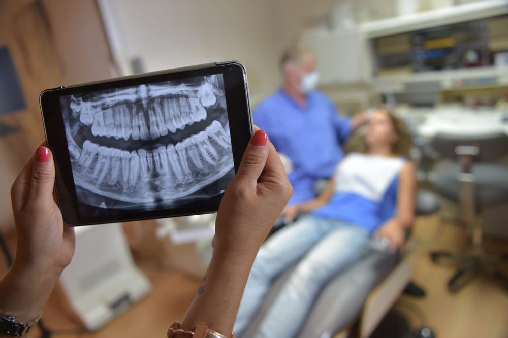
Over the course of your life, there’s a strong chance you will have to undergo a dental x-ray. They are an essential part of creating treatment plans. They can pick up tiny issues that are still invisible to the dentists’ eyes. This makes them especially useful because it is always better to place the emphasis on prevention, rather than cures.

How Do Dental X-Rays Work?
X-rays work by passing energy through soft and hard structures of your body. This energy is then absorbed by dense structures (like teeth and bone) and less so by soft structures (like your gum and decay). The balance of this energy creates the image.
Two types of x-rays are used in dentistry – intraoral and extraoral. As the names suggest, intraoral is taken from inside the mouth while extraoral is taken from the outside. The most common types used in dental practices are intraoral. These can be used to:
- Check the health of tooth roots
- Look for signs of decay and cavities
- See if the gums are suffering from periodontal disease
- Monitor the health of the jaw bone
- Check on tooth development in children and adolescence
Types of X-Rays
Bitewing
Taking their name from the shape of the x-ray machine, this is the most common form of x-ray. They show teeth above the gum line and the height of the bone between teeth. They are especially useful in detecting little spots of decay that can cause infections of the root canal. These x-rays are also used to help diagnose gum disease.
Panoramic
This machine gives dentists a view of your teeth and bones to show any irregularities. It captures the entire mouth in a single image, including the teeth, upper and lower jaws, surrounding structures and tissues. They are particularly useful in checking wisdom teeth positions, planning orthodontic treatments as well as issues with the bite. You stand inside the device, and it moves around your head to produce a panoramic image.
Periapical
This x-ray is taken to show the entire tooth including its root structure and surrounding bone structures. It offers specific images and can show infections and abscesses. It uses the same technology as the bitewing x-ray.
Dental Cone-beam CT (CBCT) Scans
Stands for dental cone beam computed tomography. It is a special type of x-ray equipment used when regular dental or facial x-rays are not sufficient. Your dentist may use this technology to produce three dimensional (3-D) images of your teeth, soft tissues, nerve pathways and bone in a single scan.
Digital X-Rays
This is a newer technology. Instead of using x-ray film, a flat electronic pad or sensor is used. The image is transferred to a computer where it can be immediately viewed and printed. It can also be stored and shared among health care providers. This type of x-ray uses half the radiation of conventional film, which is perfect for patients that are concerned about radiation. We, at Care Dental Camberwell, uses this type of latest digital radiography equipment to ensure minimal radiation exposure to all our patients.
What About Radiation?
If you have any concerns about radiation, your dentist should take these into account and plan treatments accordingly. But, there are some truths you should be aware of:
- X-rays are only performed when necessary, so you are never exposed to unnecessary radiation.
- Modern machines use very low doses. In fact, you are probably exposed to more walking down the street that you would be during the session. In fact, having two intraoral x-rays taken has been likened to walking under the sun for only 30 minutes!
- Digital x-rays produce very little radiation, and even conventional processes work so quickly that you are exposed to far less than you would have been in the past.
- A lead shield can be used while the x-ray is being taken, so only your face comes in contact with radiation. An x-ray sensor holder will be used when having the x-ray taken, so not even your fingers have to be exposed to radiation.
- The benefits of dental x-rays far outweigh its risks. The radiation exposure you are subjected to over an entire lifetime of dental treatment will be carefully considered and only recommended when necessary and the total amount should not be enough to warrant concern about your next appointment.
Dental X-Rays For Children
X-rays are entirely safe for use with children. But, your dentist will try and limit their use as much as possible. Ensuring your child follows a healthy oral health routine is the best way to limit the number of x-rays they need.
The Procedure For Dental X-Rays
Your x-ray will always be performed in the office of your dentist. In some cases, you may be sent to another specialist if you require an advanced form of radiography. The procedure should run like this:
- The protective lead apron will be fitted to your body
- Your dentist/technician will insert the x-ray sensor and holder into your mouth. You will be given instructions to guide the holder in the desire position.
- The dentist/technician will begin to image the target area
The entire procedure is pain-free and quick. Your dentist will be more than happy to discuss any fears you have, and it’s important to remember that health professionals only have your best interests at heart.
The x-ray has allowed dentistry to evolve. It allows professionals to find and treat problems early, so you don’t have to lose teeth or go through expensive and invasive interventions. It’s a vital part of holistic dentistry and something that will go a long way to securing the health of your teeth, gums, cheeks and jaws for the rest of your life.
